Going in for a cardiac positron emission tomography (PET) scan can be nerve-wracking, especially if you’ve never experienced one before. But worry not — we’re here to provide some insight into the procedure so you know what to expect. Discover how a myocardial viability study works, along with some steps you can take to prepare for the scan.
What Is a Viability Scan?
A myocardial viability PET scan measures the amount of heart muscle damage after a cardiac condition or injury, such as coronary artery disease or a heart attack. It assesses the heart muscle’s viability and the blood supply to your heart.
A myocardial viability scan uses a radioactive tracer to view the heart. This imaging can help the doctor determine if a patient requires a procedure like:
- Coronary artery bypass surgery: This surgery creates a new path for blood to flow around a fully or partially blocked coronary artery.
- Angiography: This procedure uses X-ray imaging to closely examine the blood vessels.
- Heart transplant: A cardiac or heart transplant is an operation that replaces a failing heart with a healthy one from a donor. It’s typically performed on patients with severe coronary artery disease or end-stage heart failure when other treatments have been unsuccessful.
PET viability scans differ from computed tomography (CT) scans in a couple of ways. While a CT scan produces detailed, nonmoving images of the body’s organs, tissues and bones, a PET scan provides an in-depth view of bodily tissues on a cellular level. It can help identify cancer and other abnormal activity in its earliest stages.
A CT scan is an effective diagnostic tool, but in most cases, it only shows signs of a medical condition after a disease alters the tissue or organ structure.
How Does a Nuclear Viability Study Work?
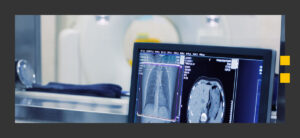
During the cardiac viability study, you’ll lie on your back on a table attached to a scanner. The radiology technologist will attach adhesive electrodes to your chest to monitor your heart’s electrical activity, then inject a radioactive tracer into the bloodstream to measure the heart’s resting blood flow.
The PET camera detects radiation from the tracer to create images. You’ll lie still with both arms overhead as the PET camera takes images of your heart.
The technologist then injects a separate radioactive tracer with fluorodeoxyglucose (FDG) and glucose. You’ll again lie still in the same position as the camera takes pictures of your heart and measures your blood sugar level. The images show how different areas of the heart use glucose, identifying which parts of the heart may have been damaged.
Cells that have been damaged or destroyed from a heart attack or heart disease use little to no glucose. Conversely, cells recovering from an injury and healthy cells use more glucose. The technologist then reviews the two sets of PET images together and sends the report to your doctor.
How to Prepare for a Myocardial Viability Study

You’ll want to follow these tips when preparing for a PET viability scan:
- Avoid strenuous activity. Exercise increases glucose in skeletal muscles, potentially affecting your scan results. Try to avoid strenuous physical activity and exercise a few days before the test.
- Go on a low-carb, sugar-free diet. You’ll likely be asked to avoid sugary, high-carb foods and beverages at least 12 hours before your scan, as these boost the body’s glucose levels. Sugar can also compromise results as PET imaging detects sugar metabolism. Instead, consume a high-protein diet of nonstarchy vegetables, hard cheeses, nuts and meat.
- Fast. You’ll need to avoid eating or drinking anything but water for 6-8 hours before the procedure. Fasting lowers your blood sugar levels, allowing the tracers to work more effectively.
- Wear proper clothing. Your technologist may ask you to change into a hospital gown. You’ll also need to remove any jewelry, as metal can interfere with the testing equipment.
- Discuss medical devices and implants with your doctor. Artificial hips and pacemakers won’t affect your PET scan results. However, you shouldn’t have a PET scan if you have any nonapproved medical devices or metal implants.
- Discuss diabetes with your doctor. If you have diabetes, fasting before your test may affect blood sugar levels. You can likely take your insulin as usual and have a small meal a few hours before your test. During the scan, you’ll be given a small amount of glucose and insulin through an intravenous (IV) line to increase glucose absorption in the heart tissue.
Are There Any Risks Associated With PET Cardiac Scans?
You may want to consider the possible side effects if you have any of these medical conditions:
- Pregnancy: You should not undergo a PET scan if you’re pregnant or think you may be pregnant. The radiation is unsafe for the developing fetus.
- Allergies: If you’re allergic to iodine, saccharin or aspartame, you may experience an allergic reaction to the radioactive tracer. The specialist may prescribe you an alternative substance.
- Fear of needles or claustrophobia: The PET scan may be uncomfortable or anxiety-inducing if you’re afraid of needles or enclosed spaces. Discuss options with your doctor to make the process more relaxing.
Because an IV will be inserted to administer the radioactive tracer, you may also experience some bruising, swelling or bleeding from the insertion site.
Benefits of a Viability Test for Your Heart
A PET viability scan is an effective way to identify heart-related conditions and determine the appropriate treatment. Your health care provider can use the scan results to help diagnose, treat or monitor a condition. Additionally, we provide the following benefits for our patients seeking myocardial viability scans here at Impression Imaging:
- Advanced technology: Our cutting-edge equipment and technology use the latest practices to provide the best care and accurate results for our patients.
- Knowledgable staff: Our team is highly trained and qualified in nuclear medicine radiology. Our professional and compassionate experts are devoted to meeting the needs of every patient. We maintain clear communication and a comfortable, efficient experience for both patients and their doctors.
- Quick turnaround: We have our patients’ results ready to send to their doctors’ offices within 12-24 hours following the scan.
- Free transportation: We provide complimentary transportation to and from our facility for anyone that needs it.
Contact Us for a Myocardial Viability Scan Near You
Whether you need a PET scan for medical care or you’re looking to learn more about the procedure, turn to Impression Imaging in southern Florida. Specializing in a variety of medical imaging services including PET scans, CT scans and more, we aim to provide every patient with exceptional and personalized care.
Whatever the reason you might require a cardiac viability study, you can trust our expert medical staff to deliver prompt services, innovative technologies and reliable, accurate results needed to diagnose and address your condition. Contact us today to learn more about viability tests for heart procedures or request an appointment.

