Dynamic imaging in nuclear medicine is an imaging technique that captures sequential images of physiological processes inside of the body. In simpler terms, dynamic imaging takes images of things like blood flow and shows them on a display processing unit.
With dynamic imaging, doctors and nuclear medicine technologists can analyze cellular progression and fluid circulation to diagnose or monitor a broad range of illnesses or conditions. Keep reading to learn more about the benefits of dynamic imaging in nuclear medicine.
What Is Dynamic Imaging?
Dynamic imaging is used in various types of imaging, including magnetic resonance imaging (MRI), computerized tomography (CT) scans, X-ray imaging and nuclear medicine. Its use in nuclear medicine involves the administration of radioactive pharmaceuticals to patients over a specific period during nuclear imaging tests such as a positron emission tomography (PET) scan. To perform dynamic imaging during a nuclear imaging test, a technologist introduces a radiopharmaceutical into the bloodstream.
Sequential still images are taken over a specific duration to analyze the flow of this substance and other fluids to a particular organ or tissue. The sequential images that dynamic images capture may be taken as quickly as every 1/10 of a second or as methodically as every 10 minutes or more per still. After the photos are taken, they can be played back rapidly to create a time-lapse movie effect.
The general purpose of dynamic imaging is to analyze the passage of blood flow through certain tissues or organs. Some particular organs that dynamic imaging is used for include the following:
- Heart
- Kidneys
- Bile ducts
- Liver
- Gallbladder
What Is Static Imaging?
While dynamic imaging is like a time-lapse movie of fluid flowing through tissues or organs, static imaging is like a snapshot of those tissues and organs. In this way, the difference between dynamic imaging and static imaging is the type of images these scans produce. Static imaging in nuclear medicine also involves the administration of radiopharmaceuticals to provide greater image contrast or clarity.
What Is Nuclear Medicine?
Nuclear medicine is a unique medical field that uses radiopharmaceuticals to evaluate various bodily functions and detect signs of disease or illness. Nuclear medicine can also identify and eradicate damaged or diseased tissue.
While both nuclear medicine and radiology use radiation to create images of bodily structures, they’re not the same type of imaging procedure. Nuclear medicine relies on internal radiation waves, meaning the images are produced from radioactive substances inside the body. In contrast, radiology produces images by applying external radiation waves through the body.
A common misconception is that MRI scans are an example of nuclear medicine. MRI scans are not nuclear medicine; MRI scans use external magnets and radio waves to produce detailed pictures of internal structures in the body on a nearby display processing unit.
Examples of nuclear imaging includes:
- Brain single-photon emission computerized tomography (SPECT) scans
- Dopamine transporter (DAT) scans
- Gallium-67 scans
- Gastric emptying scans
- Hepatobiliary scans with or without cholecystokinin (CCK), a hormone in your gastrointestinal (GI) tract
- Limited or whole body bone scans
- Liver and spleen scans
- Liver flow SPECT scans
- Lung quantification scans
- Lung ventilation-perfusion (V/Q) scans
- Multigated acquisition (MUGA) scans
- Parathyroid scans
- Renal scans with or without Lasix, a diuretic medication
- SPECT bone scans
- Thyroid uptake scans
- Triple phase bone scans
The Two Most Common Types of Imaging in Nuclear Medicine

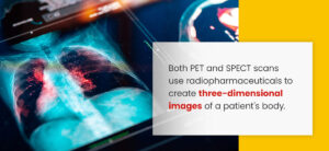
The two most common types of imaging in nuclear medicine are PET and SPECT scans. Both PET and SPECT scans use radiopharmaceuticals to create three-dimensional images of a patient’s body. The difference between these two scans is that they each use different types of radiopharmaceuticals, known as radioactive tracers.
Radioactive tracers, also known as radiotracers, consist of carrier molecules bonded to radioactive atoms that interact with specific cells, proteins or sugars in the body. PET scans use radiotracers that decay into positrons, while SPECT scans use radiotracers that contain and measure controlled amounts of gamma rays. Positrons and gamma rays produce different amounts of energy, influencing the image results.
PET scans are commonly used to detect cancer, heart problems and various brain disorders. PET scans can differentiate cancerous and noncancerous cells based on their metabolic activity. Since cancerous cells have more metabolic activity than noncancerous cells, they appear brighter on PET scans.
They’re also used to identify decreased blood flow throughout your heart. The radiopharmaceuticals produce less energy with reduced blood flow, causing those areas to appear in different colors and levels of brightness. In the brain, PET scans can detect parts of the brain that need extra glucose, which may indicate abnormalities in brain function.
SPECT scans are used for various purposes, including diagnosing and monitoring brain, heart, liver and bone disorders. Like PET scans, SPECT scans identify these issues by highlighting areas of reduced blood flow, cancer progression, bone healing and other phenomena of concern.
Benefits of Dynamic Imaging in Nuclear Medicine
The use of dynamic imaging in nuclear medicine provides many benefits to nuclear medicine technologists, doctors and patients.
For nuclear medicine technologists, dynamic imaging expands the scope of what they can test for. For doctors, dynamic imaging makes diagnostic determinations and monitoring of various illnesses more accessible. These advantages also apply to patients, as they can receive more accurate diagnoses and high-quality monitoring of their condition so they can get proper treatment.
Here are a couple of the specific benefits of dynamic imaging in nuclear medicine:
More Diagnostic Information Than Static Images
As dynamic imaging records the passage of fluid through various tissues and organs, it holds more potential for detecting information about patients’ bodies than static imaging.
Static imaging provides stills of the body, which can be helpful when assessing tissue or structural damage. However, when testing for issues concerning circulatory systems or physiological processes, static imaging has significant limitations. An example of the diagnostic information that dynamic imaging can detect is singular measurements of the blood flow around the heart’s muscles, known as myocardial blood flow.
Illuminates Ultra-Fast Phenomena
Dynamic imaging in nuclear medicine enables the illumination of ultra-fast phenomena on a cellular level. By taking sequential images, dynamic imaging can capture ultra-fast phenomena through a series of images and play it back at a slow enough rate to detect abnormalities.
Visit Impression Imaging for Imaging Services
Impression Imaging provides high-quality imaging services with state-of-the-art equipment. Whether you need an MRI, CT scan, X-ray or nuclear medicine scan, our imaging technologists can get the job done for you. If you’d like to learn more about dynamic imaging or want to schedule an appointment for a nuclear medicine scan with us, call us at (954) 580-2780 or fill out our contact form today!


